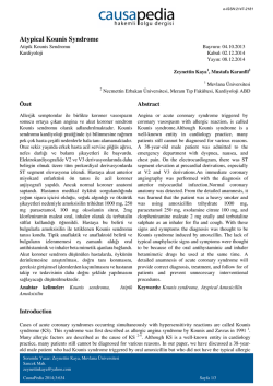
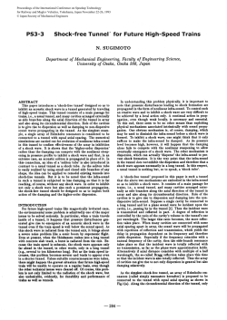
VISUAL DIAGNOSIS
A Rare Cause of Compression of the
Ulnar Nerve Neuritis: Deep Vein Thrombosis
Atif BAYRAMOGLU,1 Altan CALMASUR,2 Ayhan AKOZ,1 Murat SARITEMUR1
Deparment of Emergency, Ataturk University Faculty of Medicine, Erzurum;
1
Department of Radiology, Regional Training and Research Hospital, Erzurum
2
A 21-year-old woman presented to the emergency
department with complaint of pain in the right
elbow, and ring and pinky finger numbness. The
patient had no prior history of systemic disease.
The patient reported similar symptoms had occurred intermittently after physical exertion during the previous three months. However, the patient presented at the hospital after experiencing
the symptoms while at rest. Physical examination
identified a palpable soft mass at the beginning
of the third distal humerus and extending to the
cubital region on the inner side of the right elbow
with the consistency of the dough. The Tinel test
was positive. Other system examination findings
were normal. The patient’s laboratory findings,
complete blood count and blood chemistry tests
were normal. Magnetic resonance imaging of the
patient is shown in Figure 1 and Figure 2a, b. [see
page 130 for diagnosis]
Figure 1. An increase in brachial diameter and thrombus in fat-suppressed proton density T1-weighted MR images sequences at the
same level (*).
(a)
(b)
Figure 2. (a) An increase in brachial diameter and thrombus in axial T1-weighted MR images. Thin white
arrows: brachial vein filled within thrombus. *: m. pronator teres, **: m. brakialis, black triangles: a. brachialis, curved white arrow: v. basilica, thick white arrow: v. cephalica. (b) An increase in brachial diameter and thrombus in fat-suppressed proton density in the axial image at the same level with figure 2a.
Submitted: August 21, 2013 Accepted: October 31, 2013 Published online: January 20, 2014
Correspondence: Dr. Atıf Bayramoğlu. Ataturk Universitesi Tıp Fakultesi Kampus, 25240 Erzurum, Turkey.
e-mail: [email protected]
Turk J Emerg Med 2014;14(3):97 [130]
doi: 10.5505/1304.7361.2014.59319
97
130
VISUAL DIAGNOSIS
[see page 97]
DIAGNOSIS: Cubital Tunnel Syndrome
Cubital tunnel syndrome (CuTS) is the most common
form of entrapment of the ulnar nerve and the second most common nerve compression syndrome of
the upper extremities, the most common being carpal tunnel syndrome. The incidence is 24.7 cases per
100,000 person-years; men are affected twice as often
as women. Clinical findings include paraesthesia and
tingling in the lateral 2 fingers, pain related to the elbow joint, and in severe cases, weakness or atrophy of
the intrinsic muscles of the hand.[1-3] Soft tissue masses such as ganglia, lipomas, fibrolipomas, and epidermoid cysts can compress the ulnar nerve as it lies
in the condylar groove or within the cubital tunnel.
However, it was not reported that the ulnar nerve was
wrapped and compressed by the DVT mass. In this respect, the present case has the distinction of being
unique. We present the first case of CuTS due to DVT.
Magnetic resonance imaging of the patient detect-
Turk J Emerg Med 2014;14(3):97 [130]
ed an acute phase deep vein thrombosis (DVT) that
could not be compressed, causing an increase in the
lumen diameter in the brachial vein and in the right
elbow (Figure 1, 2). After cardiovascular surgery,
consulting physicians initiated medical treatment
with enoxaparine (150 anti-Xa IU/kg, once-daily).
Control returned to the patient and the patient was
discharged. The patient’s symptoms completely resolved three months later.
References
1. Assmus H, Antoniadis G, Bischoff C, Hoffmann R, Martini AK, Preissler P, et al. Cubital tunnel syndrome - a
review and management guidelines. Cent Eur Neurosurg 2011;72:90-8. CrossRef
2. Fullbier L, Renz B. Operative treatment of cubital tunnel syndrome. Aktuel Neurol 2011;38:298-302.
3. McPherson SA, Meals RA. Cubital tunnel syndrome.
Orthop Clin North Am 1992;23:111-23.
doi: 10.5505/1304.7361.2014.59319
© Copyright 2026 Paperzz